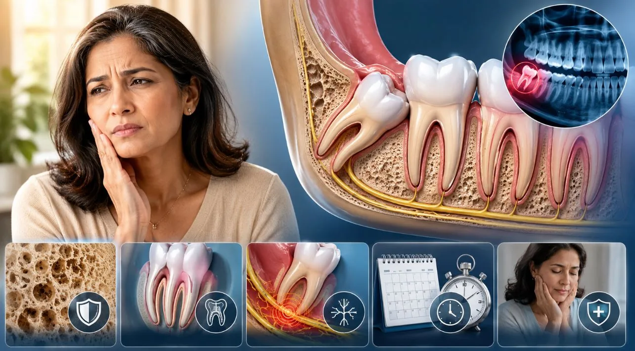
When watching is the right call
The other half of the conversation, the half that gets less airtime, is that not every retained third molar in a forty-year-old needs to come out. A fully erupted, properly aligned, functionally occluding third molar that the patient can clean and that has no decay, no pocketing, no radiographic concerns, and no symptom history is, for many people, a tooth that can be left alone for life. The American Association of Oral and Maxillofacial Surgeons has been clear that prophylactic extraction of asymptomatic, disease-free third molars in older patients is not automatically indicated, and the Cochrane reviews on the topic have repeatedly concluded that the evidence does not support a one-size-fits-all extraction policy.
What watching really means is monitoring, not ignoring. It means a panoramic radiograph every few years, attentive probing of the periodontal pockets around the tooth, a careful look at the distal surface of the second molar for early decay, and a low threshold for reconsideration the moment something changes. It also means an honest conversation about what the patient's mouth will look like in a decade or two. A third molar that is fine at forty-five but is harder to clean than its neighbours might be a different story at sixty.
Two specific cases where watching is often the wise call:
- The fully bony impaction in an asymptomatic adult. A wisdom tooth that is completely covered by bone, with no communication with the mouth, has no path for bacteria to reach it. The risk of leaving such a tooth in a forty-five-year-old has to be weighed against a meaningfully higher surgical risk to remove it — particularly if the roots are anatomically close to the nerve canal. Many of these are best left alone, monitored, and addressed only if something changes.
- The patient with significant medical comorbidity. A poorly controlled diabetic in their fifties with no symptoms from a quiet third molar is not a patient who needs to be in a surgical chair this month. The benefit of extraction is theoretical; the cost is concrete. Stabilising the underlying condition first, or accepting that the tooth will stay, are both legitimate decisions.
Wisdom-tooth surgery after forty is meaningfully harder than the same procedure at twenty — denser bone, mature root anatomy often near the inferior alveolar nerve, slower healing — but routinely done with good outcomes by surgeons who handle these cases regularly. The right framing is matching surgical complexity to the experience of the operator, not deferring to age alone.
Paraphrased editorial summary of the AAOMS White Paper on third molar management1 and the Cochrane review on retention versus removal2
What the surgery actually involves at forty-plus
The single most important pre-operative step in this population is three-dimensional imaging. A conventional panoramic X-ray is a flat picture of a curved anatomy and can underestimate or overstate the relationship between a third molar root and the inferior alveolar nerve. A cone-beam CT scan, which most modern oral surgery practices have on site, shows the relationship in three dimensions and allows the surgeon to plan the osteotomy and the order of root sectioning before touching tissue. If you are over forty and being told that your lower third molars need to come out, asking explicitly whether a cone-beam CT will be done first is a reasonable question. In some cases — clear separation, simple anatomy — it may not change the plan. In many cases it will.
The surgery itself is most often done under local anaesthesia with sedation, either intravenous sedation or oral. General anaesthesia is reserved for difficult cases or strong patient preference. The duration of the procedure varies enormously: a fully erupted upper wisdom tooth in an adult with healthy bone can come out in under five minutes. A lower full-bony impaction with curved roots near the nerve in a fifty-year-old can take forty-five minutes per side. A good surgeon will tell you which of those scenarios you are in before you are sedated, not after.
Sectioning the tooth — cutting it into pieces with a fine bur so that each piece can be released along the path of least resistance — is the central technique. It is far gentler than trying to elevate a whole intact tooth out of mature bone. The price is more bone reduction and more heat, which is why irrigation, careful drilling, and a surgeon who works deliberately rather than quickly matter so much.
Recovery, told as a realistic timeline
Recovery after a difficult lower third molar extraction at forty-five is not the same as recovery at twenty-two. Plan for that honestly. The classical advice — ice for the first day, soft food, no straws, no smoking — all still applies. What is different is how long each of these phases lasts.
Day zero (the day of surgery)
The first eight hours are about haemostasis and swelling control. Bite firmly on the gauze for the time you are told to. Ice the cheek over the surgical site, twenty minutes on and twenty minutes off. Eat nothing solid; nothing hot; nothing through a straw. Take the analgesics on schedule, not when pain starts, because pain that is allowed to build is harder to bring back down. Most patients do not need opioids past the first twenty-four hours, but a short prescription is often sensible after a difficult extraction.
Days one to three
Swelling peaks somewhere on day two or three and is normal — sometimes startlingly so. Bruising can spread down the neck. Trismus (difficulty opening the jaw) is common and not a sign of anything wrong. Soft foods, gentle warm-salt-water rinses starting on day two, scrupulous attention to keeping the rest of the mouth clean while leaving the surgical site alone. This is the window in which dry socket, if it is going to happen, will declare itself, classically as a deep throbbing pain that develops several days after surgery and is not well controlled by the usual analgesics. If that happens, call the surgeon — it is treatable and not an emergency, but it does not resolve on its own quickly.
Days four to seven
The worst of the swelling subsides. Most patients are back to a normal work routine by the end of week one, though anyone whose work is physically demanding or whose week involves public speaking may want to plan an extra day. Sutures, if non-dissolvable, come out somewhere in this window. You will still feel tender, still need to chew on the opposite side, still need to avoid the temptation to probe the socket with your tongue.
Weeks two to six
The socket gradually fills in with soft tissue and then with bone. Food trapping in the healing socket is normal and frustrating; a curved-tip syringe with warm salt water, used gently, is the right tool. By week six most patients have forgotten the surgery happened. A few will continue to have intermittent altered sensation if the nerve was disturbed; in most of those cases the sensation continues to improve over months. Permanent change is rare but possible — which is exactly why the consent conversation matters.
Three honest questions to ask the surgeon before the surgery
- How many of these difficult extractions do you do a month? This is the single most useful question and the one patients rarely ask. Volume tracks closely with outcome for technique-sensitive surgery. You are looking for a number measured in weeks, not years.
- Have you looked at a three-dimensional image of this tooth, and what does it show about the nerve? If the answer is "we will use the panoramic," for a lower third molar in a patient over forty, that is a fair conversation to push on. For uncomplicated upper teeth or anatomically clear lower teeth, it may genuinely not change the plan. For anything ambiguous, it should.
- What is the realistic recovery for someone my age and health? A surgeon who tells you it will be "just like the kids" without asking about your overall health, medications, smoking history, and how physically demanding your week is, is not being thoughtful. A good answer is specific to you.
The bottom line
Wisdom teeth removal after forty is meaningfully harder than wisdom teeth removal at twenty, and it is also routinely done, with very good results, by surgeons who do these cases all the time. The right framing is not "I should have done this earlier" or "I shouldn't be doing this now." The right framing is: this tooth either needs to come out, or it doesn't, and if it does, this is a surgery that benefits from imaging, an experienced operator, and a respectful approach to a body that has aged.
For some patients the answer is straightforward and the surgery is straightforward and the recovery is uneventful and the question fades. For others the right answer is to leave a quiet tooth quiet. The conversation that gets you to the right answer is worth having properly, with someone who is paying attention to the specific facts of your mouth and your health rather than reciting a default.
If you are weighing this decision now, the most useful first step is usually a panoramic radiograph and a thoughtful second opinion from a surgeon who handles difficult third molars routinely. Most general dentists know when a case is in their scope and when it isn't; an experienced oral surgeon will often spend the consultation talking you
out of unnecessary surgery as readily as into necessary surgery.
Find a clinic near you on Smyleee or
browse dentists by specialty to start that conversation with someone whose work you can actually evaluate.
Frequently asked questions
- Is wisdom-tooth removal at 40 risky?
Riskier than at 20, but not dangerous in trained hands. Inferior alveolar nerve disturbance rates are 2–10× higher depending on the imaging picture; dry socket rates climb; recovery is slower. Most cases are routinely done with good outcomes, particularly when imaging is thorough (a cone-beam CT for difficult lower impactions is standard), the surgeon handles these cases routinely, and the post-operative plan is realistic about a longer recovery than a 22-year-old would have.
- Can I leave my wisdom teeth alone at 40?
Sometimes, yes. A fully erupted, functional, cleanable wisdom tooth with no decay, no periodontal pocketing, no symptoms, and no radiographic concerns can be left in place lifelong. The American Association of Oral and Maxillofacial Surgeons has been clear that prophylactic extraction of asymptomatic disease-free third molars in older patients is not automatically indicated. Watchful waiting with periodic imaging is a defensible position for the right patient.
- How long is recovery at 40+?
Plan for 7–10 days of meaningful symptoms versus the 3–5 days a 22-year-old might experience. Peak swelling at 48–72 hours, return to desk work usually by day 4–5 for most patients, full bone healing of the socket over months rather than weeks. Patients with physically demanding work, public-speaking roles, or significant comorbidities should plan more time off than they would have at a younger age.
- What imaging do I need before surgery?
A panoramic X-ray for screening. For difficult lower impactions — particularly anything where the panoramic suggests root proximity to the inferior alveolar nerve canal — a cone-beam CT is standard of care. The CBCT shows the 3D relationship between roots and nerve that flat radiographs cannot, and changes surgical planning when it identifies high-risk anatomy. Asking explicitly whether CBCT will be ordered is reasonable for any over-40 lower impaction.
- Should I see an oral surgeon or general dentist?
For an over-40 wisdom tooth with any complexity, an oral and maxillofacial surgeon who handles difficult third molars routinely. Volume matters substantially at this difficulty level — a surgeon doing 10 difficult cases per week has a different outcome curve than one doing 10 per year. A simple upper third molar in healthy bone can be handled by a general dentist with surgical training; a full bony impaction near the nerve canal is specialist work.
- Does insurance cover wisdom-tooth extraction at any age?
Generally yes for medically indicated extractions — symptomatic, decayed, damaging the adjacent molar, or with cyst formation. Coverage for prophylactic extraction of asymptomatic teeth varies by plan and is often limited or contested in older adults given the lower threshold for medical necessity. Verify with the practice's billing office before surgery; both dental and medical insurance may be involved for complex cases.
Sources & further reading
- American Association of Oral and Maxillofacial Surgeons. "Management of Third Molar Teeth." AAOMS White Paper.
- Ghaeminia H, Nienhuijs MEL, Toedtling V, et al. "Surgical removal versus retention for the management of asymptomatic disease-free impacted wisdom teeth." Cochrane Database of Systematic Reviews. 2020;5:CD003879.
- Renton T, Yilmaz Z. "Profiling of patients presenting with posttraumatic neuropathy of the trigeminal nerve." Journal of Orofacial Pain. 2011;25(4):333–344.
- Bui CH, Seldin EB, Dodson TB. "Types, frequencies, and risk factors for complications after third molar extraction." Journal of Oral and Maxillofacial Surgery. 2003;61(12):1379–1389.
- Dodson TB, Susarla SM. "Impacted wisdom teeth." BMJ Clinical Evidence. 2014;2014:1302.
- Kandasamy S, Rinchuse DJ, Rinchuse DJ. "The wisdom behind third molar extractions." Australian Dental Journal. 2009;54(4):284–292.
How we wrote this
This piece draws on the peer-reviewed sources and authoritative guidelines listed below, and where appropriate also on patient-facing materials from the relevant professional bodies and the National Institutes of Health. Each substantive claim links to its source via the inline footnote next to it — click any number to jump to the citation. Where the evidence is genuinely uncertain or contested, the text says so rather than presenting one position as settled. We do not accept clinic, device, or pharmaceutical sponsorship for the content of editorial articles.
This article was last medically reviewed in June 2026 by the Smyleee Medical Advisory Board. We update when significant new evidence emerges or when published guidelines change. If you have feedback on a specific claim or believe an updated source warrants inclusion, please contact our editorial team.
Editorial note. This article is provided for general informational purposes and is not a substitute for individualised medical or dental advice. It reflects the evidence and clinical reasoning current at time of publication; specific decisions about extraction, surgery, anaesthesia, or post-operative care should be made in consultation with a licensed dentist or oral and maxillofacial surgeon who has assessed your own imaging, medical history, and goals. Reviewed by the Smyleee Medical Advisory Board.